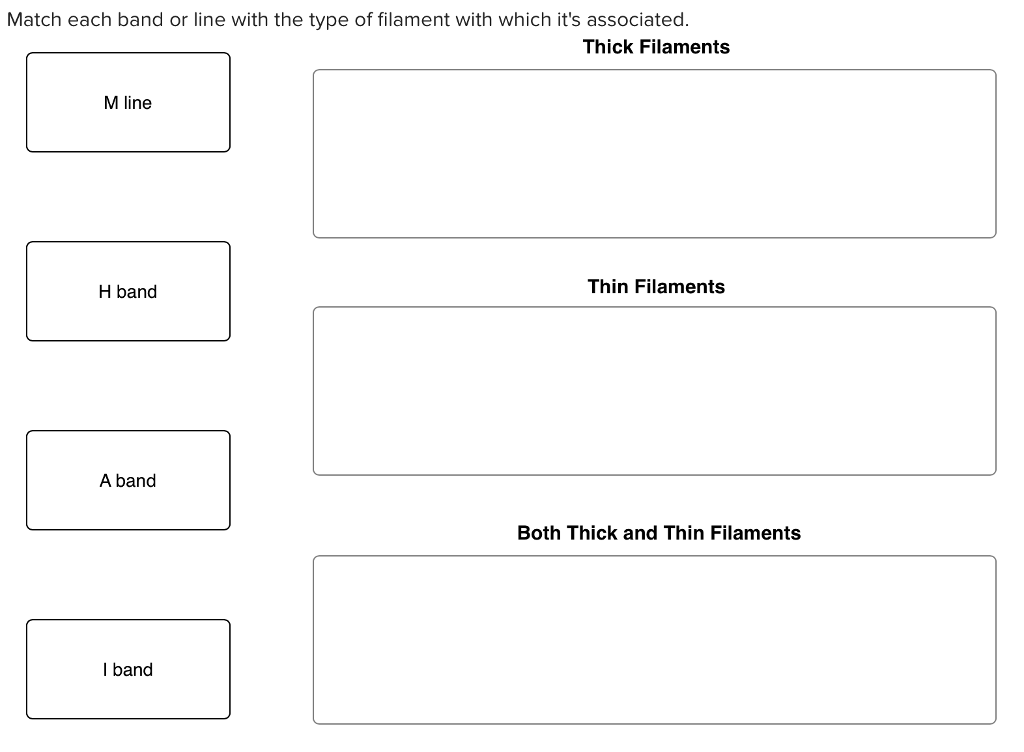
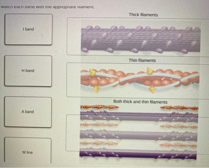
Match Each Band With the Appropriate Filament.
The M-line is located in the mid of Z-lines containing myomesin. Thick filaments I band Thin filaments H band Both thick and thin filaments A band M line.

A P Module 6 Flashcards Quizlet
Branch of a motor neuron is.

. Match each protein with the appropriate filament. Semi-permeable membrane made of phospholipids that acts as a barrier between inside and outside of the cell. Check the running extruder temperature is match with the temperature of the filament extrusion needed.
I band light Thin filament made of actin A band dark H zone Thick and thin filaments slide past each other 0 000 I band light Thick filament made of myosin Thick When a nerve impulse arrives at the neuromuscular junction acetylcholine is released stimulating an action potential in the sarcolemma. The H zone in the middle of the A band is a little lighter in color because the thin filaments do not extend into this region. The diagram above shows part a myofibril called a sarcomere.
During contraction the H-zone I-band the distance between Z-lines and the distance between M-lines all become smaller. Blocking of myosin head. The two I-bands contain a thin filament while the thick filaments are not too far away.
The sliding filament contraction occurs in the sarcomere region. Sarcomeres repeat themselves over and over along the length of the myofibril. Contains DNA and controls the cells activity.
Each myosin is composed of multiple units of meromyosin which has two important parts- a globular head known as heavy meromyosin with a short arm and a tail known as light meromyosinThe head and arms project at regular distance and angle from each other from the surface of myosin filament and are known as the cross armThe head bears binding sites for. During muscular contraction the length of thick filament or thin filament does not change. Key Points For Sliding Filament Theory.
Chapter 9 Muscles and Muscle Tissue 279 9 B. Part A Drag the labels to the appropriate location in the figure. Within the A band is the H zone which is the area composed only of thick myosin.
The mechanism of contraction is the binding of myosin to actin forming cross-bridges that generate filament movement Figure 1. When a sarcomere contract two adjacent Z lines come closer to each other. The center of zone H has a vertical line called line M where accessory proteins hold together the thick filaments.
Recently the crystal. H zone - It is the center of sarcomere in which there is no overlap between actin and myosin in relaxed state. If not keep trying to set a appropriate temperature.
Match the column I with column II. However the A bands size remains constant during contraction. This gave researchers an idea of myosins central location.
Place a single word into each sentence to make it correct then arrange the sentences into a logical paragraph order to describe the process of contraction of a sarcomere. The input of the device is internally matched to 50 Ω across 27 to 35 GHz. Sealing the filament in a dry storage can effectively prevent it brittle and crack cased by absorbing moisture from the air for a long term.
Match each protein with the appropriate filament. The sliding filament theory of muscle contraction was developed to fit the differences observed in the named bands on the sarcomere at different degrees of muscle contraction and relaxation. The A band is dark because of the thicker mysoin filaments as well as overlap with the actin filaments.
Each skeletal muscle is a discrete organ made up of several kinds of tissues. A sarcomere is a contractile unit of skeletal muscle that is divided. The main components of the histology of a sarcomere are summarized below.
Double membrane that surrounds the nucleus and has pores to allow some molecules like RNA out. Thick filament zone composed of myosin proteins. __C__ Sarcomere __B__ Myofibril __A__ Muscle Fiber __E__ Actin Thin Filament __D__ Myosin Thick Filament Directions.
Region of muscle fiber containing only thin filament. The Z-lines are responsible for the striped nature. Match each description with the appropriate term.
Reset Help Z line H band Zone of overlap Thick filament M line Sarcomere Thin filament I band. The fluid cytoskeleton and organelles inside a cell. The thin filaments extend into the A band toward the M-line and overlap with regions of the thick filament.
Actin and myosin overlaps each other forming cross bridge. Filament model beginning with 91 Overview of muscle types special characteristics and functions WHY THIS MATTERS. This is the smallest unit of skeletal muscle that can contract.
When muscle is at rest the overlapping of actin filament to the myosin head is. Match the Organelle to Its Function. 0203 Muscle Contraction Assignment Before you begin please take 10 minutes to watch Crash Courses fun and ex-tremely helpful video on the sliding filament theory.
Main protein found in thick filaments of muscle. PAs have been designed to optimize performance across 27 to 31 or. Region of muscle fiber that extends the full length of the thick filament.
The sequences of muscle contraction explained by sliding filament theory are as follows. Please label the following with the appropriate letter. As most S-Band radars will only use a portion of this band the PA output match may be optimized for either output power or PAE across the intended operating band.
The cross bridge is active only when myosin head attached like hook to the actin filament. Thick filaments Myosin Light chains Actin Thin filaments Troponin Active sites Tropomyosin Neither thick nor thin filaments Titin match each oana with the appropriate thiament. The A band has a higher content of thick myosin filament as expected by the areas rigidity.
The A band is the area in the center of the sarcomere where thick and thin filaments overlap. A band or anisotropic band is the region where both thick filaments and thin filaments are present. Intermediate filaments IFs commonly have structural elements of a central α-helical coiled-coil domain consisting of coil 1a coil 1b coil 2 and their flanking linkers.
These filaments slide in and out between each other to form a muscle contraction hence called the sliding filament theory. This central region of band A seems slightly lighter than the rest of band A and is called zone H. Skeletal muscle fibers predominate but blood vessels nerve fibers and substantial amounts of.
Weve got the study and writing resources you need for your assignments. 2 How do I store my filament. The A-band contains both thick and thin filaments and is the center of the sarcomere that spans the H zone.
Science Biology QA Library Part A Drag the labels to the appropriate location in the figure. A sarcomere consists of different bands that are as follows - Z line - it is the line between two adjacent sarcomeres.
Lab Practical 2 Flashcards Quizlet
Solved Match Each Band Or Line With The Type Of Filament Chegg Com
Solved Match Each Protein With The Appropriate Filament Chegg Com
No comments for "Match Each Band With the Appropriate Filament."
Post a Comment